Astrocyte Highways: The Secret Garden of the Central Nervous System
Anna Jagielska, Mingyu Yang (HST MEMP PhD student), Krystyn J. Van Vliet
MIT Department of Materials Science and Engineering
The Koch Institute Image Awards celebrate the extraordinary visuals that are produced through life sciences and biomedical research at MIT. This year, one of the awardees include an image from the Van Vliet Lab (Professor Krystyn Van Vliet, Department of Materials Science and Engineering, MIT). Also sharing in the image award are Anna Jagielska and Mingyu Yang, an HST Medical Engineering and Medical Physics (MEMP) PhD student. More info on the awards and other winners, here.
Read on for more about the winning image.
How would you describe the subject of this image to a layperson?
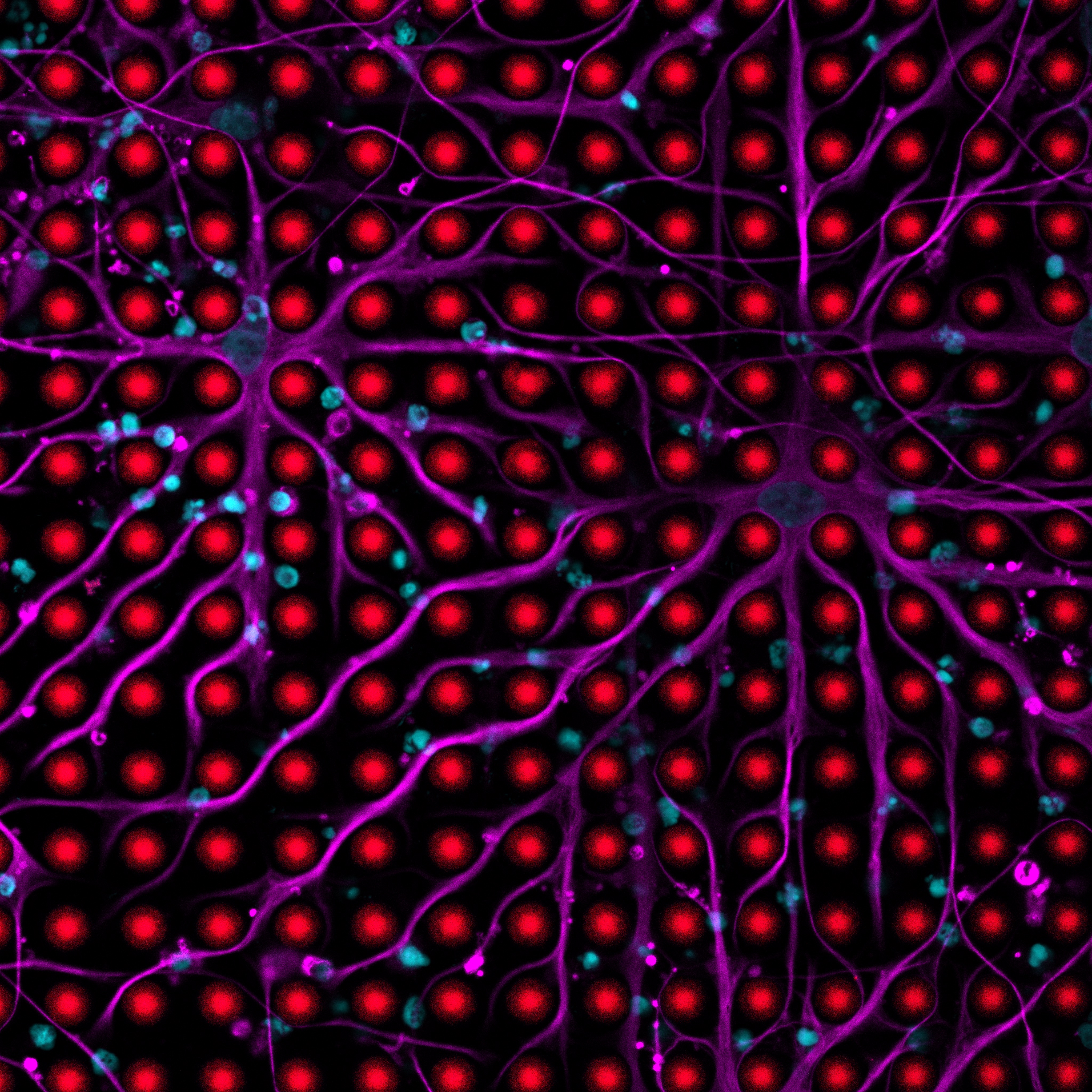
These images show glial cells (human and rodent oligodendrocytes, astrocytes and
microglia) interacting with engineered synthetic 3D-printed Artificial Axons, mimicking
biological axons of neurons. Artificial Axons, the first fully controllable synthetic axon
mimics, were developed at the Van Vliet lab by A. Jagielska and others to study vital
process of myelination and the interaction of glial cells with axons, in the tunable in vitro
settings. Artificial Axons are currently used for drug discovery of compounds to promote
myelin repair in Multiple Sclerosis and to study myelin disorders in Alzheimer’s disease.
Why did you take this image? What were you trying to learn?
These images were taken to see at high magnification how glial cells engage with
axons. In case of oligodendrocytes, our goal was to demonstrate that these cells can
wrap myelin membrane on the 3D-printed mimics of axons, as it happens in the Central
Nervous System (CNS).
How does this image fit into the larger goals of your research?
These images provided a validation that the Artificial Axons platform can indeed mimic interactions
observed in the CNS. Specifically, this is the first synthetic platform that allows for
imaging and direct quantification of myelin wrapping in the high throughput setup,
allowing us to use it for the discovery of compounds to stimulate myelin repair in MS.
In one sentence, please tell us what is most exciting about this image.
These are the first images of the of intimate interactions of glia with axon mimics
proving that our axon mimics can be used to model myelination, which is extremely hard
to study in vivo.
Technical Details
What type(s) of microscope/equipment and techniques/preparations were used to
create the image?
Fluoview 3000, Olympus. Immunostaining with fluorescently tagged antibodies.
What is the scale/magnification of the image?
Images were taken with varied magnifications, using 20x and 30x lenses and zoom 1x,
6x or 8x
Is there anything else you’d like to tell us about your submission?
These are first ever images of myelination on 3D-printed axon mimics!